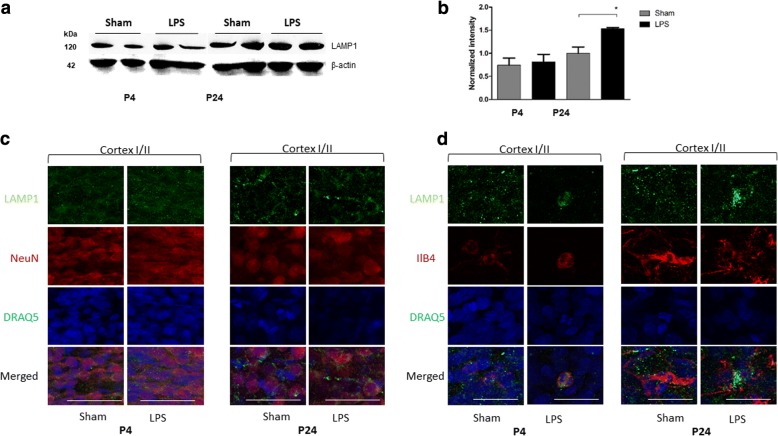
Fig. 1.
LAMP-1 protein level was increased 21 days after LPS exposure. LAMP-1 protein levels were measured by Western blot in the brain homogenates of rat pups injected with LPS (n = 8) and Sham (n = 8) 1 day (P4) and 21 days (P24) after injection, *p < 0.05 (a, b). LAMP-1 distribution in upper layers of somatosensory cortex (I–II) in neurons (NeuN-positive cells) (c) and microglia (ILB4-positive cells) (d) of LPS and Sham animals at P4 and P24 was analyzed by immunofluorescent confocal microscopy. Cortex I–II, cortical layers according to [64]

