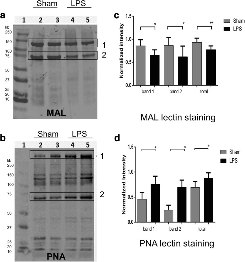
Fig. 7.

21 days after LPS injection, rat brain tissue glycoproteins show reduced staining for MAL-II lectin and increased staining for PNA lectin consistent with their desialylation. a, b Representative glycoprotein patterns of brain tissue homogenates of Sham (lanes 2–3) and LPS-injected (lanes 4–5) rat pups at P24 after injection. Following SDS-PAGE, blots were stained with biotinylated peanut agglutinin (PNA) lectin, specific for Gal-GalNAc residues and biotinylated Maackia amurensis II (MAL II) lectin, specific for α2–3-linked sialic acids. The bands showing apparent difference in the intensity between LPS and Sham samples are boxed. c, d Intensity of lectin-positive bands normalized to the level of total protein. For statistical analysis, five animals from each group were used; *p < 0.05, **p < 0.01
