Fig. 8.

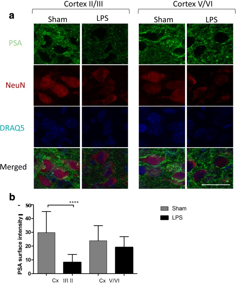
21 days after LPS exposure, upper cortical neurons show reduced PSA immunostaining. a Neurons with decreased PSA immunostaining are detected in the upper somatosensory cortex (layers II–III) of LPS-injected rat pups at P24 (n = 3) after injections as compared to Sham animals (n = 3). PSA, green; neuronal marker NeuN, red. Bar graphs (b) show average PSA immunostaining intensity in cortical neurons counted for nine adjacent 0.0042-mm2 sections of upper and lower layers of the somatosensory cortex. Bar represents 20 μm. ****p < 0.0001. n.s., non-significant, Cx, cortex; I–VI, cortical layers according to [64]
