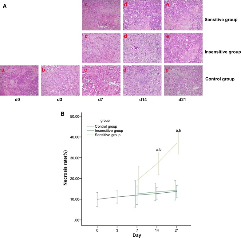
Fig. 9.
Effect of DTX on tumor necrosis in rat EOCs. Microscopic pictures (HRF × 200) on day 0 (a), day 3 (b), day 7 (c), day 14 (d), and day 21 (e) of the histopathology of EOC specimens (A), which are demonstrated as spotty or patchy necrosis scattering in the tumor, were analyzed. Tumor necrosis was increased in the sensitive group but showed no remarkable change in the insensitive or control group. On days 14 and 21, tumor necrosis (B) was significantly different among the three groups, except for that in the insensitive vs. control group. p < 0.05, a: sensitive vs. control; b: sensitive vs. insensitive

