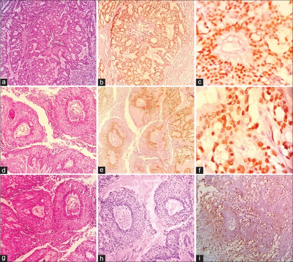
Figure 1.
Photomicrographs of ameloblastic carcinoma (a) Proliferating odontogenic islands (H and E, ×100). (b) Diffuse and high SOX-2 nuclear staining of basal and parabasal layers (IHC ×100). (c) High SOX-2 nuclear staining of tumor cells (IHC ×200). (d) Odontogenic island with central necrosis (H and E, ×100). (e) Diffuse and high OCT-4 nuclear staining of basal and parabasal layers (IHC ×100). (f) High OCT-4 nuclear staining of tumor cells (IHC ×400). (g) Odontogenic islands with central necrosis and retraction artifact (H and E, ×100). (h) Negative staining for OCT-4 in tumor islands (IHC ×100). (i) Low membrane expression for CD44 in the tumor islands (IHC ×400)

