Figure 4.

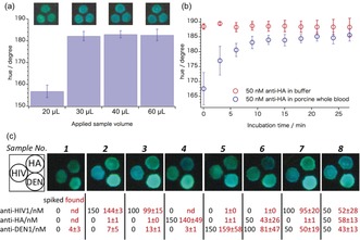
3D‐μPADs applied to antibody assays in whole blood: a) hue values obtained from variable amounts of porcine whole blood spiked with 50 nm anti‐HA applied to an anti‐HA targeting 3D‐μPAD; data recorded 21 min after sample application. Photos above the graph show the colorimetric response as recorded by the camera. b) Time‐dependent bioluminescence emission hue values recorded from anti‐HA‐LUMABS modified 3D‐μPADs after application of 20 μL aqueous anti‐HA samples or 30 μL of anti‐HA spiked porcine whole blood. Error bars (a,b) indicate the SD for readouts from three different signal detection areas on one single device. c) Photos of signal detection areas of 3D‐μPADs responsive to multiple antibodies recorded 21 min after application of antibody‐spiked porcine whole blood; spiked and found antibody concentrations are indicated in black and red, respectively; confidence intervals represent SD for triplicate readouts from three separate devices.
