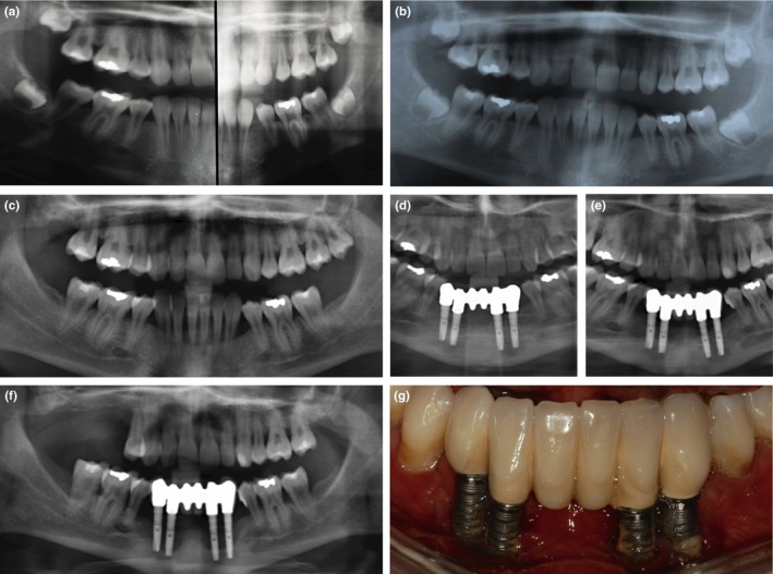
Figure 1.

Periodontal and peri‐implant bone loss in individual 1. (a) Splitted orthopantomogram at age 14 years: At the end of orthodontic treatment, no periodontal bone loss is evident. (b) Orthopantomogram at age 16 years and (c) age 18 years: Periodontal bone loss with rapid progression is clearly visible in the mandibular anterior region. (d) Implant placement after bone augmentation, and prosthetic reconstruction at age 24 years. (e) Peri‐implant bone loss 3 years, and (f) 5 years after implantation. (g) Rapid progressing peri‐implantitis was clinically characterized by the absence of pocketing, but receding gums and exposed implant threads
