
Figure 3.
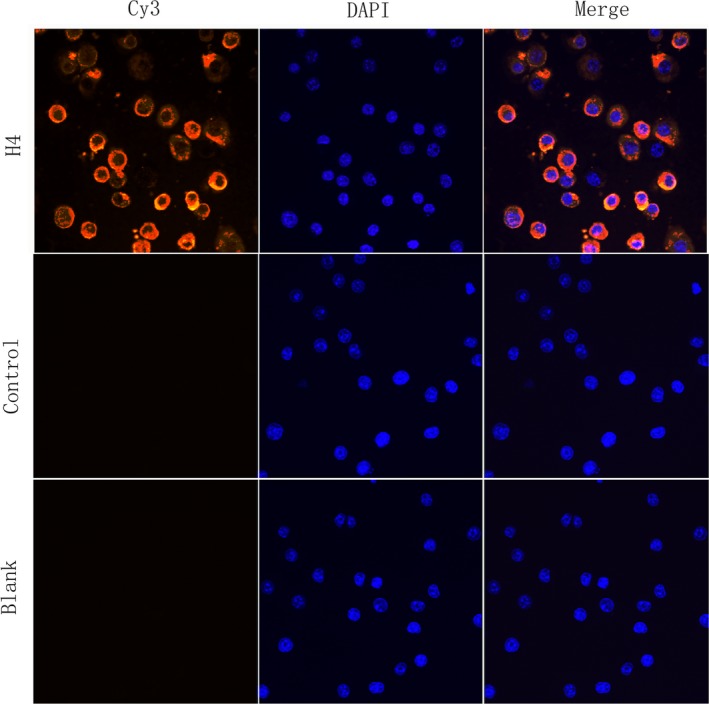
rTgH4 binds to murine macrophages. The nuclei of the macrophages were stained by DAPI (blue). Cy3‐conjugated secondary antibody was used to visualize the rTgH4 protein (red). Merged, overlapping the blue channels with red channels. No red fluorescence was noted in the PBS blank group or the pET‐32a vector protein control group.