
Figure 7.
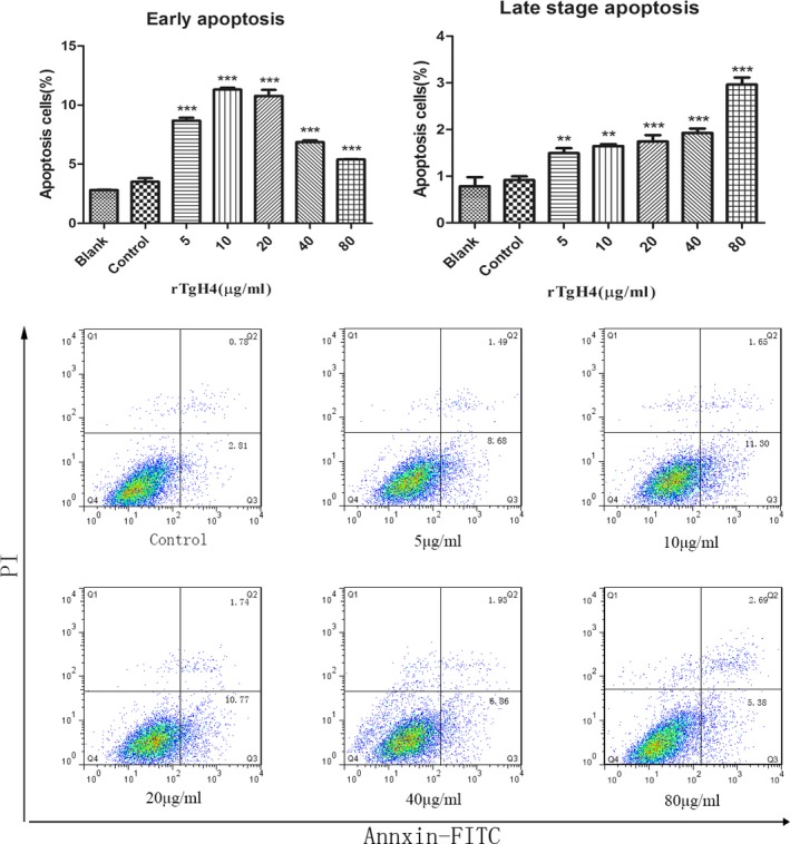
rTgH4 protein induced the apoptosis of the murine macrophages. After incubation with rTgH4, apoptosis was quantified by flow cytometry utilizing an Annexin V‐FITC kit. In the blank group, the cells were treated with PBS, and in the control group, the cells were treated with pET‐32a vector protein. The data were indicative of three individual experiments (**p < 0.01 and ***p < 0.001).