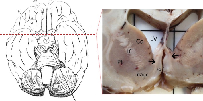
Figure 1.

An illustration of the diagonal band (d–d′) in the human brain seen from the inferior surface, with the optic tract retracted, as described in Broca's monograph in 1888 12 (left). Coronal section at a level approximately 1.5 cm anterior to the mammillary bodies (red dashed line) revealed the extent of the diagonal band (black arrows) on both sides of the hemisphere, medial to the basal ganglia (right). Cd, caudate; IC, internal capsule; LV, lateral ventricle; nAcc, nucleus accumbens; PO, paraolfactory area; Pt, putamen.
