
Figure 4.
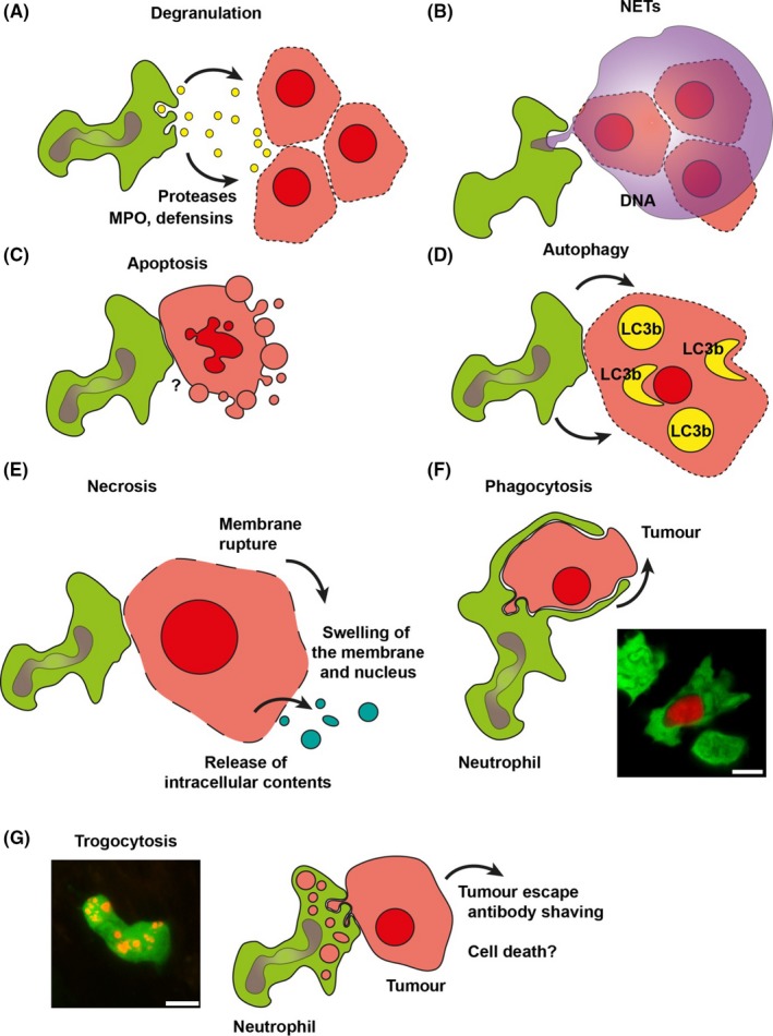
Mechanisms of antibody‐dependent elimination of tumour cells by neutrophils. The mechanisms by which neutrophils kill antibody‐opsonized tumour cells are ill‐understood and sometimes controversial. The mode(s) of action depends among others on the type of antibody, target cell and (density of) tumour epitope. This large number of variables might explain the variety of distinct effector mechanism that has been described in the literature. This includes tumour cell killing via A, degranulation; B, release of neutrophil extracellular traps (NETs); C, apoptosis; D, autophagic cell death; E, necrosis; F, phagocytosis and G, uptake of tumour‐derived membrane resembling trogocytosis. The outcome of the latter process is controversial. Trogocytosis may contribute to killing, but it has also been shown that this process by contrast protects tumour cells by shaving off tumour antigen‐antibody complexes. Pictures in f and g were acquired with a Leica TCS SP8 STED 3X super‐resolution microscope equipped with white light laser and Hybrid photodetectors. Neutrophils (green) and rituximab opsonized human B‐CLL (red) in the spleen of a LysM‐EGFP mice were recorded with a voxel size of 0.38 × 0.38 × 1 μm, a 40× numerical aperture (NA) 1.3 oil immersion objective and a pixel dwell time of 1.96 μs