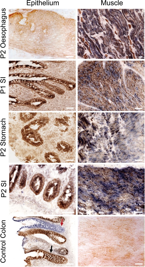
Figure 2.

Deficient COX activity in post‐mitotic smooth muscle but normal COX activity in mitotic epithelium of the alimentary canal in patients with inherited m.3243A>G. The left panel shows the COX‐normal epithelium that is labelled brown in the SI of patient 1, and the oesophagus, the stomach, and the SI of patient 2, while the right panel manifests the blue COX‐deficient muscle fibres in these tissues. The control panel shows the COX‐normal epithelium (black arrow) and smooth muscle from a normal individual who also has crypts with defective COX activity (red arrow), due to accumulated somatic mtDNA mutations during ageing. Scale bar = 50 μm.
