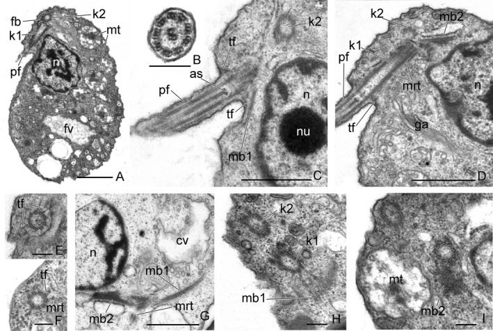
Figure 3.

The general view, anterior end of the cell, and some flagellar root structures of Aquavolon hoantrani. A. The longitudinal section of the cell. B. Cross‐section of the flagellum. C‐D. Longitudinal sections of the anterior end of the cell. E‐H. Relative arrangement of the kinetosomes. I. Some microtubular bands near the nucleus. as, axosome; cv, contractile vacuole; fv, food vacuole; ga, Golgi apparatus; k1, kinetosome 1; k2, kinetosome 2; mb1, microtubular band 1; mb2, microtubular band 2; mt, mitochondria; mrt, microtubules; n, nucleus; nu, nucleolus; pf, posterior flagellum; tf, transition fibers. Scale bar: (A, C, D, I) 1 μm, (B, E – H) 0.2 μm.
