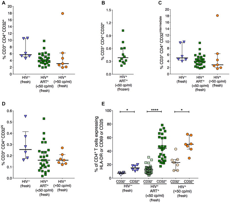
Fig. 1. CD32+ CD4+ T cells are enriched with activated cells.
Freshly isolated peripheral blood mononuclear cells (PBMCs) from HIV-controls, HIV+ individuals with VL <50 copies (cp)/ml, and HIV+ individuals with VL >50 copies/ml were stained for CD32, CD69, HLA-DR, and CD25 on CD4+ T cells. (A) Percentage of total CD32+ within CD4+ T cells. (B) Percentage of total CD32+ CD4+ T cells in cryopreserved PBMCs from HIV+ individuals with VL <50 copies/ml. (C) Percentage of CD32intermediate within CD4+ T cells. (D) Percentage of total CD32hi within CD4+ T cells. (E) Percentage of cells expressing at least one of the activation markers human leukocyte antigen-DR (HLA-DR), CD69, and CD25 on CD32+ CD4+ T cells and CD32− CD4+ T cells. Lines and error bars represent median and IQR, respectively. All statistical comparisons were performed using two-tailed Wilcoxon rank tests. n = 6 for HIV− controls, n = 27 for HIV+ ART+ (<50 copies/ml) individuals, and n = 7 for HIV+ (>50 copies/ml) individuals. *P < 0.05 and ****P < 0.0001.

