
Figure 6. Transient decrease in p53 protein levels is sufficient to trigger cell cycle re-entry in arrested cells.
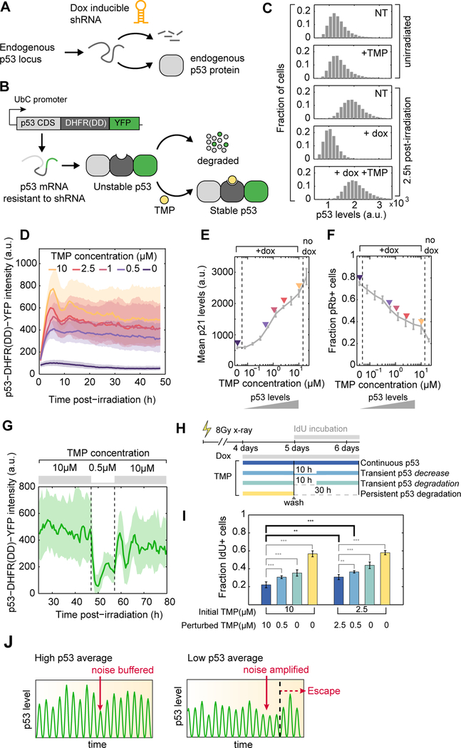
(A) A dox-inducible shRNA allows depletion of endogenous p53 protein.
(B) Exogenous expression of a p53 protein fused to a DHFR destabilizing domain is stable only in the presence of TMP. The p53-DHFR(DD)-YFP mRNA is resistant to the inducible shRNA described in (A).
(C) Distributions of p53 protein levels in single cells, as quantified by immunofluorescence. TMP- dependent stabilization of p53-DHFR(DD)-YFP effectively rescues shRNA-mediated depletion of endogenous p53 in response to DNA damage.
(D) p53-DHFR(DD)-YFP levels can be gradually modulated by addition of increasing doses of TMP. Medians +/− interquartile ranges of single cell trajectories are shown (n > 100 cells per condition).
(E, F) Cells received 2Gy xray and were treated with increasing concentrations of TMP in the presence of dox. Average p21 levels and the fraction of pRb positive cells were quantified 2 days post-irradiation using immunofluorescence (n = 3 biological replicates).
(G) Transient decrease in TMP concentration reversibly reduced p53 levels in individual cells. Medians +/− interquartile ranges of single cell trajectories are shown (n = 67 cells).
(H) Schematic of experimental design to test the effect of transient disruption of p53 signaling on cell fate. Cells received 8Gy xray and were treated with dox and TMP 4 days post-irradiation for 24h. Media was washed and replaced with original TMP concentration (continuous p53), a low TMP concentration for 10h (transient decrease), no TMP (transient degradation) for 10h or no TMP until the end of experiment (persistent degradation). Media was supplemented with IdU to quantify the proportion of cells that go through S-phase post-wash.
(I) Proportion of cells that incorporate IdU as a function of TMP treatment schedule and base TMP concentration (n = 6 biological replicates, ** p < 0.01, *** p < 0.001).
(J) Heterogeneity in p53 pulse amplitude defines a permissive state within which temporal fluctuations in signaling drive cell cycle transitions. In a cell with high amplitude of p53 pulses the noise is buffered. In a cell with lower averaged p53 pulse amplitude the noise is amplified and results in escape from arrest.