
Figure 4.
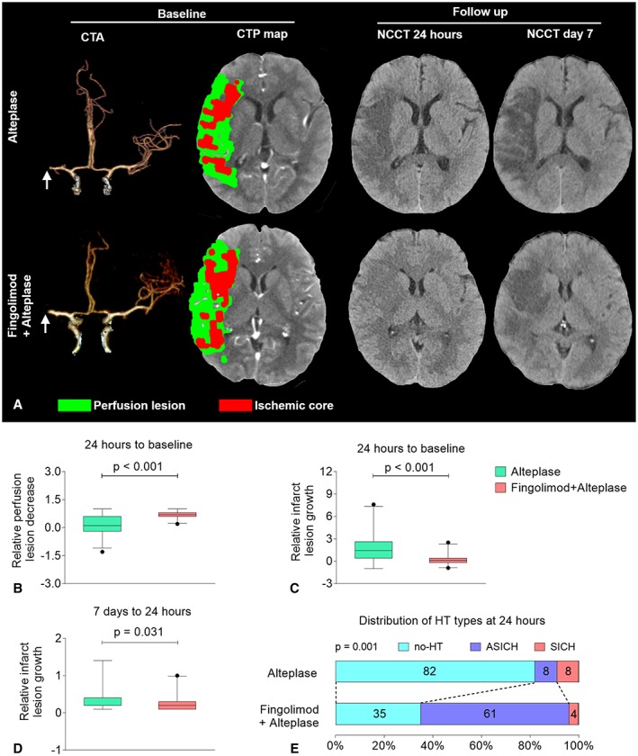
Fingolimod decreased perfusion lesions and restrained infarct growth at 24 hours among patients with intravenous alteplase treatment. (A) Representative multimodal computed tomographic (CT) scans of patients with alteplase treatment (upper panel) and fingolimod plus alteplase treatment (lower panel). At baseline, although the site of artery occlusions (arrows) and the mismatch status were the same, striking differences were evident at follow‐up. The progression of infarct volume was restrained in fingolimod‐treated patients. (B) The decrease in perfusion lesion at 24 hours. The relative perfusion lesion decrease was defined as 1 − (perfusion lesion volume at 24 hours / perfusion lesion volume at baseline). Positive values for the relative perfusion lesion decrease rate indicate improvement. (C) The growth in the infarct lesion at 24 hours. The relative infarct lesion growth was defined as (infarct lesion volume of noncontrast CT [NCCT] at 24 hours / ischemic core volume of CT perfusion [CTP] at baseline) − 1. Negative values for the relative infarct lesion growth rate indicate improvement. (D) The growth of the infarct lesion from day 7 to 24 hours. The relative infarct lesion growth was defined as (infarct lesion volume of NCCT at day 7 / infarct volume of NCCT at 24 hours) − 1. Negative values for the relative infarct lesion growth rate indicate improvement. The horizontal line inside each box indicates the median, the top and bottom of the box indicate the interquartile range, the bars indicate the 5th and 95th percentiles, and the circle indicates an outlier. (E) The distribution of hemorrhagic transformation (HT) types at 24 hours. The numbers in the bars are percentages of patients who had the same type. Symptomatic intracranial hemorrhage (SICH) was defined as large parenchymal hematoma combined with a significant clinical deterioration of ≥4 points on the National Institutes of Health Stroke Scale (NIHSS). Asymptomatic intracranial hemorrhage (ASICH) was defined as a small parenchymal hematoma combined with clinical deterioration of <4 points on the NIHSS. CTA = CT angiography.