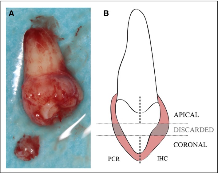
Figure 2.

Illustration of the sampling process. (A) Dental follicle during the sampling procedure in which the coronal portion has been sectioned. (B) The dental follicle was dissected into three different fractions. The middle portion was discarded, and the coronal and apical portions were independently fixed. Subsequently, each of the apical and coronal parts was divided in half, with one half being used for gene‐expression analyses and the other half being used for immunofluorescence. IHC, immunohistochemistry.
