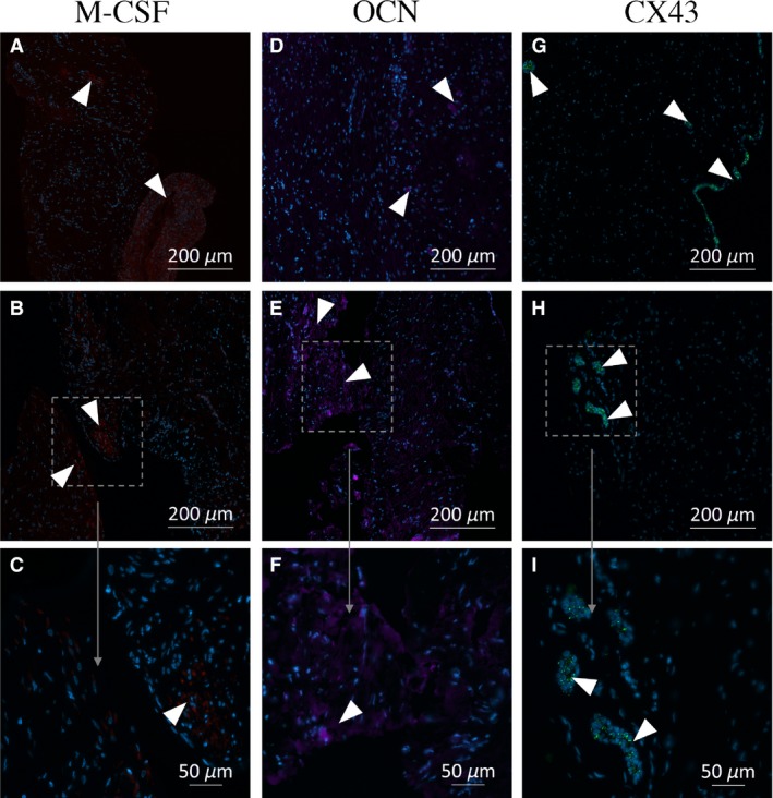
Figure 5.

Immunofluorescence photomicrographs of osteocalcin (OCN), gap‐junction protein, alpha 1, 43 kDa (CX43), and colony‐stimulating factor 1 (CSF‐1) expression in sections of dental follicle. The localizations of CSF‐1 (red), OCN (purple), and CX43 (green) are shown in representative images of sections of the human dental follicle obtained from different patients. No obvious spatial pattern of expression of the proteins is discernible. Staining with antibodies directed against bone morphogenetic protein 2 (BMP‐2) and RANKL was negative for all the samples. Positive areas are enlarged for a more detailed evaluation. The sections examined are from the following patients and portions of the follicle: (A) Patient 1, coronal; (B, C) Patient 3, apical; (D) Patient 3, apical; (E, F) Patient 1, coronal; (G) Patient 5, apical; (H, I) Patient 2, coronal.
