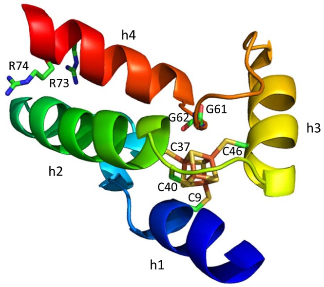
Figure 4.

The Structure of WhiB1 from Mycobacterium tuberculosis (Kudhair et al., 2017). Labelled are the four conserved Cysteine residues (C9, C37, C40 and C46), two Glycines (G61 and G62) that are part of the conserved G(V/I)WGG turn and the C‐terminal Arginine residues (R73 and R74) that are proposed to interact with DNA in M. tuberculosis. Helices are labelled (h1‐h4). PDB ID:5oay.
