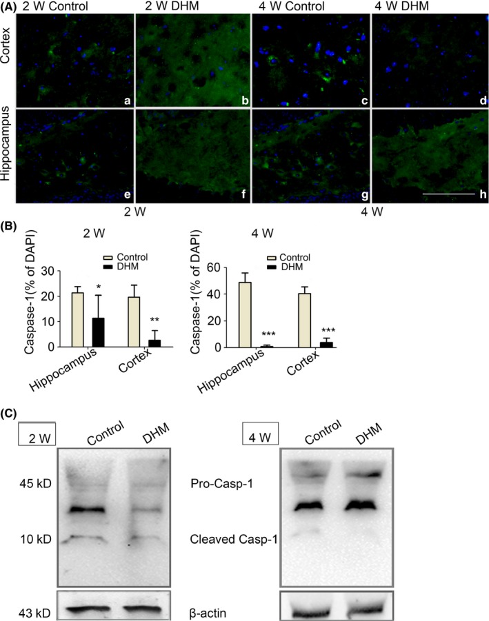
Figure 3.

Caspase‐1 (Casp1) protein reduce after dihydromyricetin (DHM) treated in APP/PS1 mice. A‐B, Caspase‐1 immunostaining revealed decreases expression of Caspase‐1 in the hippocampus and cortex of DHM‐treated mice, as compared with control groups mice. Scale bars: 40 μm. (*P < .05, **P < .01 vs Control group at 2 weeks, ***P < .001 vs control group at 4 weeks). C, Two bands of 45 and 10 kDa were detected on Western blots using the Caspase‐1 antibody. Given this 10 kDa molecular weight, this protein is probably activated Caspase‐1. Caspase‐1 was almost detectable following DHM treated but was markedly in APP/PS1 control mice. β‐actin was used as the loading control
