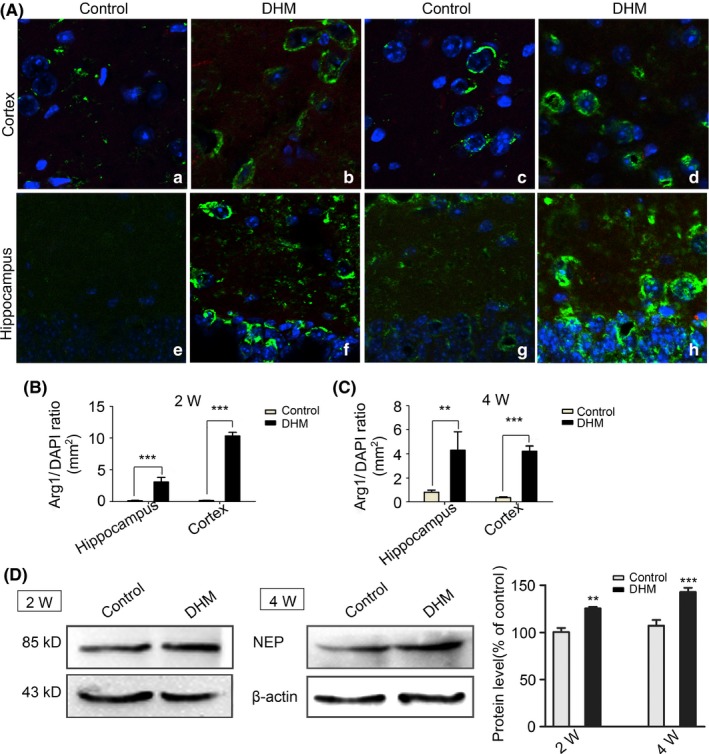
Figure 8.

M2 staining in APP/PS1 mice treated by dihydromyricetin (DHM). A, M2 immunostaining of brain sections from APP/PS1 animal show that the number of arginase‐1‐positive cells (green) increased in DHM‐treated groups compared with control groups. Scale bars: 50 μm; B, C, Bar graphs showing that the number of arginase‐1‐positive cells significantly increased in the DHM‐treated groups (**P < .01, ***P < .001 vs control groups). D, Western blot shows that the expression of NEP were up‐regulation in 2 and 4 weeks DHM administration groups compared with their control groups. *P < .05, **P < .01, ***P < .001 vs control
