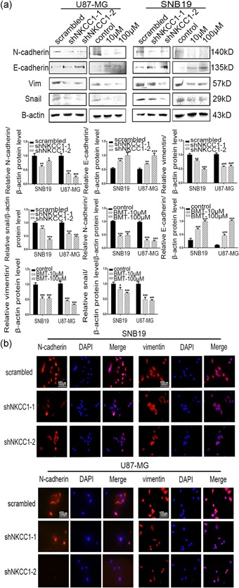
Figure 4.

The downregulation of NKCC1 reversed the epithelial–mesenchymal transition. (a) Western blotting showing the decreased protein levels of mesenchymal markers (N‐cadherin, vimentin, and snail) and the increased protein levels of an epithelial marker (E‐cadherin) after knockdown of NKCC1. The same effect was observed after treatment with bumetanide after 24 hr. β‐actin was used as a positive control. (b) Immunofluorescence staining showing the same outcomes as for NKCC1 in U87 and SNB19 cells after shRNA treatment and bumetanide treatment. The scale bar corresponds to 100 μm (*p < 0.05; **p < 0.01; ***p < 0.001). NKCC1: sodium‐potassium‐chloride cotransporter 1 [Color figure can be viewed at wileyonlinelibrary.com]
