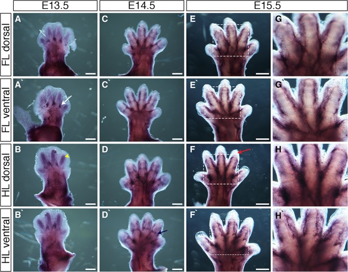
Figure 4.

Expression of Cadherin5 (Cdh5) during mouse limb development. A–H`: Whole‐mount in situ hybridization for Cdh5 at E13.5 (A–B`), E14.5 (C–D`), and E15.5 (E–F`) in dorsal forelimbs (A, C, E), ventral forelimbs (A`,C`,E`), dorsal hindlimbs (B,D,F), and ventral hindlimbs (B`,D`,F`). Strong regions of expression are seen at the digit base, between digits (A` white arrow). Blood vessels are visible in regions of developing cartilage at E13.5 (B yellow arrowhead). Bands of expression across presumptive joint sites are visible at E14.5 (D navy arrow). Avascular distal mesoderm is indicated (F` red arrow). G–H`: Higher magnification of regions highlighted in white dashed boxes in E–F`, respectively. Scale bars = 500 µm.
