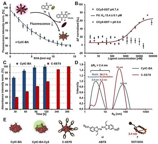
Figure 3.

A) Characterization of CytC‐BA by Alizarin Red S assay for BA group determination revealed 11 BA groups. B) Microscale thermophoresis of CCy5‐SST under pH 7.4 for binding affinity determination of SST‐SHA 7 towards CytC‐BA and protein release via SST shell cleavage by acidification. C) Colorimetric assay using ABTS vi to screen bioactivity of C‐SST8 in comparison to the free protein CytC‐BA. D) DLS results of CytC‐BA and C‐SST8, which were both incubated in PB pH 7.4, representatively shown for 90° measurement (analyses with other angles are displayed in the supporting information). E) Structures of CytC‐BA (BA‐functionalized CytC), CytC‐BA‐Cy5 (Cy5‐labelled CytC‐BA), C‐SST8 (8 equivalents of SST‐SHA 7 assembled on CytC‐BA), ABTS vi and visualization of dimension of SST‐SHA 7, which was simulated by the software Molecular Operating Environment.
