
Fig. 2.
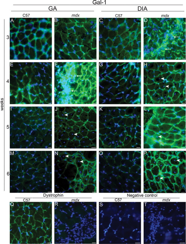
The mdx mice exhibit increased expression and extracellular localization of Gal-1 in the gastrocnemius (GA) and diaphragm (DIA) muscles during the degeneration wave. Analyzed muscles are indicated at the top: GA (A, B, E, F, I, J, M, N, Q to T) and DIA (C, D, G, H, K, L, O, P), from C57BL/6 (C57) and mdx, placed side by side. Ages are indicated at the left: 3 weeks (A–D), 4 weeks (E–H, Q–T), 5 weeks (I–L), and 6 weeks (M–P). Muscular sections were labeled using anti-Gal-1 (A–P), anti-dystrophin (Q and R), and a secondary antibody conjugated to Alexa-488. Areas containing high density of cells (DAPI staining) show more intense labeling of Gal-1 (arrows in D and F). Nonperipherally nucleated myofibers, detected only in mdx muscles and mainly in the GA, are indicated by arrowheads in the GA (J, N) and in the DIA (H, L, P). Sarcolemmal dystrophin immunostaining confirmed wt and mdx phenotype (representative images in Q and R). Negative control (absence of primary antibody) is showed in S and T. Nuclei are stained with DAPI. Bar = 10 μm.