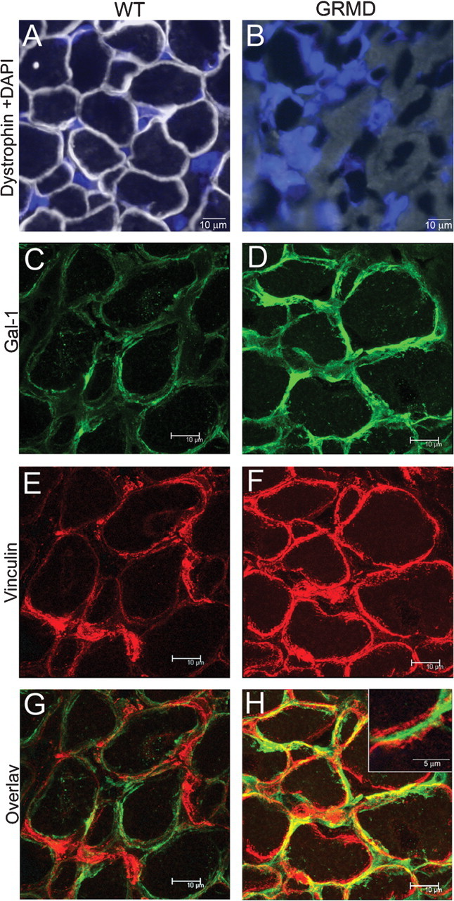
Fig. 7.

Localization of Gal-1 in GRMD femoral biceps. (A–B) Dystrophin staining in wt (A) or GRMD (B) femoral biceps. Nuclei are stained with DAPI. (C and D) Gal-1 staining of wt (C) or GRMD (D) femoral biceps sections. (E and F) Vinculin staining of wt (E) or GRMD (F) femoral biceps. (G and H) Overlay image of wt (G) or GRMD (H) for Gal-1 and vinculin. The region of tissue in which Gal-1 is localized mainly in the extracellular compartment (G, inset).
