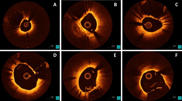
Figure 3.

OCT images of patients who had undergone SKS deployment previously. These are representative images from five different patients. For consistency, all images are taken in the LMS in the LMS‐LAD stent, with the neocarina visible between that stent and the LMS‐Cx stent. There is a continuous coverage of tissue between the struts of the neocarina in panels A‐D. Panels E and F taken from the same patient; the diaphragm is unfenestrated at the neocarina but is fenestrated towards the ostium. An OCT video pullback in SKS is viewable online. SKS barrels are visible from 20 s
