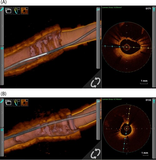
Figure 4.

OCT study of a LMS SKS case recorded 4 months post‐PCI. Panel A is taken from the LMS‐LAD limb and Panel B from the LMS‐LCX limb. The middle third of each 3D cut‐away demonstrates the neo‐carina or ‘diaphragm’, a sheet of endothelium which covers the stent adjoining the two limbs in the LMS section. The catheter is seen in the right third and the LAD (A) and LCX (B) segments are in the left third. The cross‐sectional OCT view (taken at the mid‐LMS point) shows the adjacent barrel
