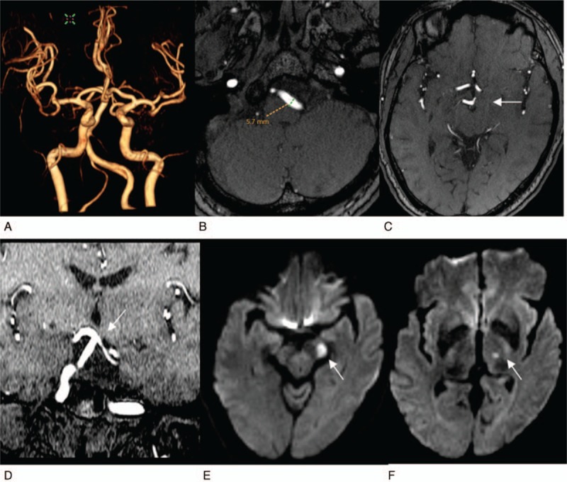
Figure 1.

It reveals a high bifurcation of the basilar artery complicated with infarction in the blood supply area of the posterior cerebral artery. The patient was a 55-year-old woman. A, 3D VR MRA reconstruction image of cerebral vessels, from the anterior-to-posterior view. The vertebrobasilar artery significantly deviates to right, showing an S shape (grade 2), and has a high bifurcation. B, C, Cross-sectional TOF images. (B) It reveals that the diameter of the left vertebral artery is 5.7 mm, (C) it reveals that the basilar artery bifurcation reaches the level of third ventricle (white arrow), at grade 3. D, TOF reconstruction imaging of coronary surface confirms that the basilar artery bifurcation reaches the lower edge of the third ventricle (white arrow) and is slightly compressed and changed. E, F, Cross-sectional images of DWI, respectively, reveal fresh lacunar cerebral infarctions of left cerebral peduncle and left thalamus (white arrows).
