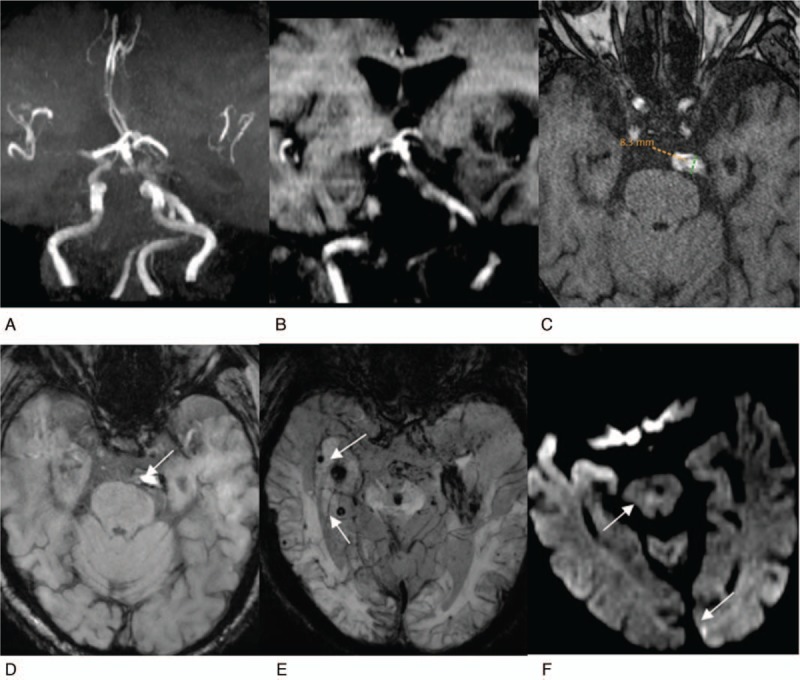
Figure 2.

Reveals a high bifurcation of the basilar artery complicated with atherosclerosis and posterior circulation infarction. The patient is 85-year-old man. A, Reconstruction MRA image of maximum density projection of cerebral vessels, from the anterior-to-posterior view. The cerebral artery presents with atherosclerotic changes, the vertebrobasilar artery significantly deviates to right, showing an S shape, reaching the left CP corner area (grade 3). B, TOF reconstruction image of the coronal surface, the basilar artery bifurcation reaches the level of the middle of the third ventricle (grade 3). C, Cross-sectional TOF image. The basilar artery signals are uneven, suggesting mural macro thrombi and atherosclerosis, with a diameter of 8.3 mm. D, E, Cross-sectional SWI images. D, It reveals low mural signal, suggesting mural thrombi (white arrow). E, It reveals the scattered distribution of “black spots” on the bilateral temporal occipital lobe of the brain stem (white arrow), suggesting micro hemorrhagic foci. F, Cross-sectional DWI image, suggesting fresh lacunar cerebral infarction of brainstem and left occipital lobe (white arrow).
