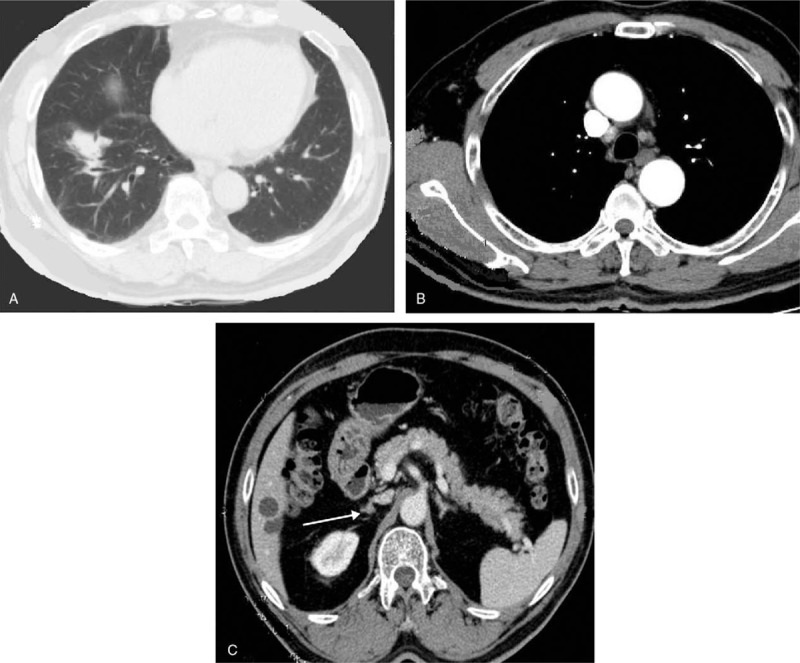
Figure 5.

In March 2016, the disease progress was evaluated by CT, which indicated lung lesions (A) and mediastinal and bilateral paratracheal lymph node (B) enlargement, and the metastatic site (C) stays the same all the time. CT = computed tomography.
