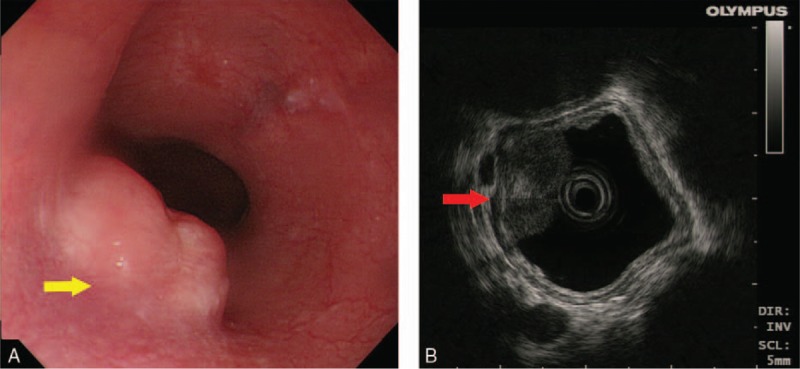
Figure 1.

Endoscopy and endoscopic ultrasound image. (A) Endoscopy showed a 1.55cm × 0.65 cm hemispherical neoplasm with a smooth surface of the middle thoracic esophagus (25 cm from incisor, yellow arrow). (B) The tumor arose from muscularis mucosae under endoscopic ultrasound (red arrow).
