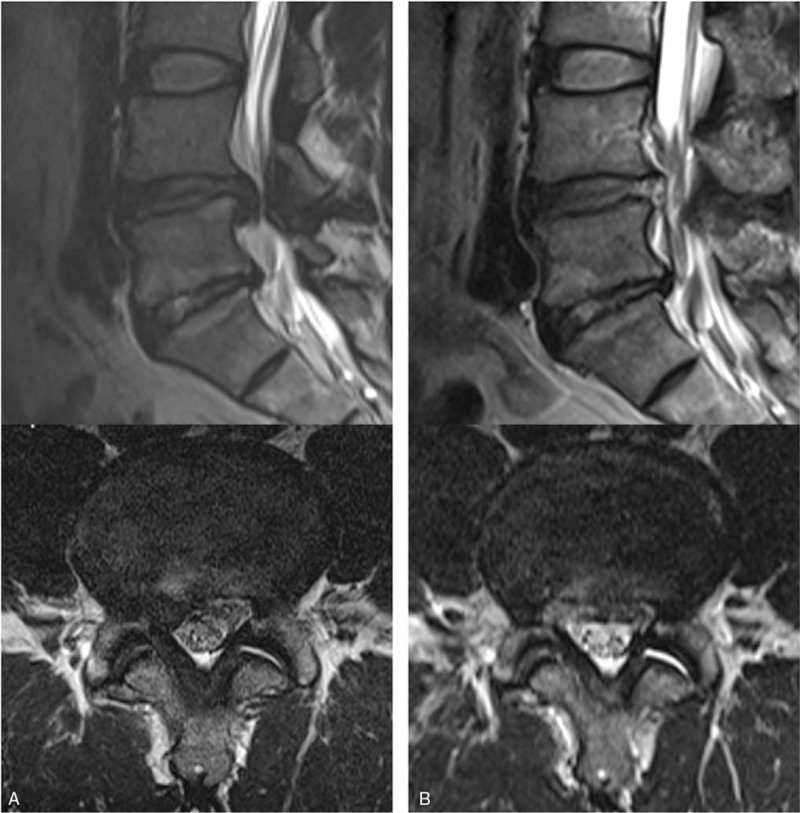
Figure 2.

Illustrated case of a 44-year-old male patient with an excellent postoperative outcome. A. Preoperative MRI showing extruded disc herniation at the right L4-5 level. B. Postoperative MRI showing complete epidural decompression after selective removal of the herniated disc. MRI = magnetic resonance images.
