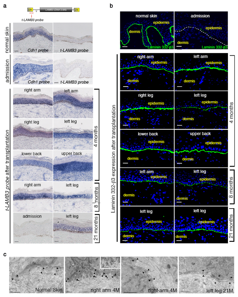
Figure 2. Restoration of a normal epidermal-dermal junction.
Skin sections were prepared from normal skin, patient’ affected (admission) and transgenic skin at 4, 8 and 21 months follow-up. a, In situ hybridization was performed using a transgene-specific probe (t-LAMB3) on 10-μm-thick sections. E-cadherin-specific probe (Cdh1) was used as a control. Scale bars, 40 μm. b, Immunofluorescence of laminin 332-β3 was performed with 6F12 moAbs on 7-μm-thick sections. DAPI (blue) marks nuclei. Dotted line marks the epidermal-dermal junction. Scale bars, 20 μm. c, Electron-microscopy was performed on 70-nm-thick skin sections. A regular basement membrane (arrows) and normal hemidesmosomes (arrowheads, higher magnification in the inset) are evident in patient’ transgenic skin. Scale bars, 1 μm.

