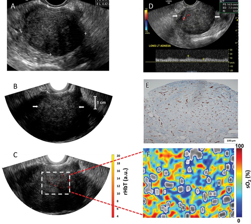
Figure 7:

Images in a 61-year-old woman after menopause with a left ovarian mass (patient 4). A, Pelvic US shows a solid, homogeneous, hypoechoic left ovarian mass with areas of posterior acoustic shadowing. B, Transvaginal US image and, C, coregistered photoacoustic tomography and pulse-echo US image shows scattered vascular components of relative total hemoglobin (rHbT) of left ovary (arrows in B) of 3.51 arbitrary units (a.u.). D, Color Doppler and spectral analysis show minimal internal blood flow (arrows). E, CD31 immunostaining in the sutured area shows dense stroma with scattered vascular components. F, Oxygen saturation (sO2) map of higher mean sO2 of 53.2%. Histopathologic examination showed a benign fibrothecoma.
