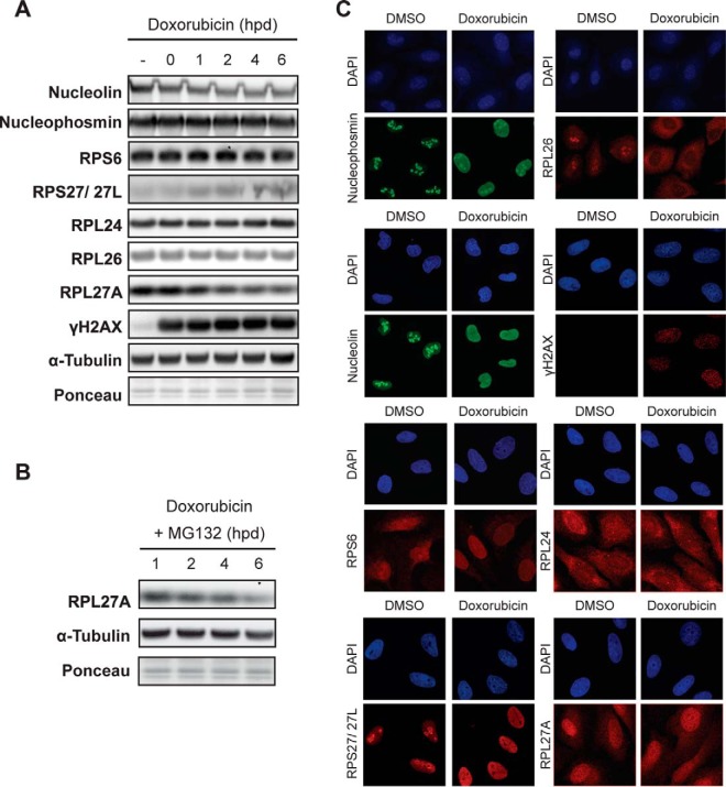
Fig. 5.
Expression level and localization of nucleolar and ribosomal proteins in response to DNA damage. (A) Expression level of ribosomal and nucleolar proteins after DNA damage. U2OS cells were synchronized in G2 and treated with a pulse of doxorubicin for 1 h. Cells were harvested at the indicated hours post-damage (hpd), and the expression of several proteins was analyzed with the indicated antibodies. Tubulin and ponceau S were used as loading controls. Both in (A) and (C), γH2AX was used as a marker for DNA damage. (B) Same as in (A) but with the inclusion of MG132 after the damaging insult. (C) Cellular localization of ribosomal and nucleolar proteins after DNA damage. U2OS cells were synchronized in G2, fixed 2 h after doxorubicin pulse and stained with the indicated antibodies using immunofluorescence. Cells were counterstained with DAPI to show the nuclei.

