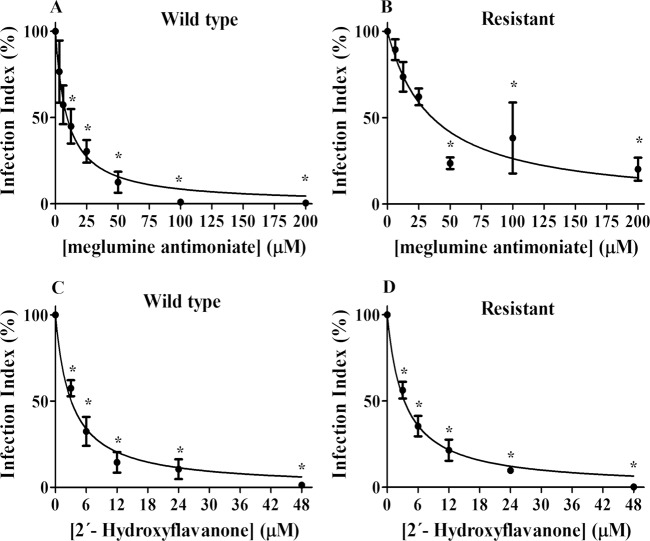
Fig 3. Effect of 2HF and meglumine antimoniate on L. amazonensis-infected macrophages.
Macrophages were infected with wild-type or antimony-resistant L. amazonensis promastigotes at 37°C and 5% CO2. After 3 hours of infection, the remaining promastigotes were removed. After 18 hours, the infected macrophages were incubated in the absence or presence of increasing concentrations of 2HF (3–48 μM) or meglumine antimoniate (3.125–200 μM) for 72 hours. The infection index was determined using light microscopy. At least 200 macrophages were counted on each coverslip in duplicate. The values shown represent the mean ± standard error of three independent experiments. In the control samples (absence of 2HF), a similar volume of vehicle (0.2% DMSO) was added to the cells. Panel A and B: Wild-type and antimony-resistant cells, respectively, treated with meglumine antimoniate; Panel C and D: Wild-type and antimony-resistant, respectively, treated with 2HF. The values are presented as the mean ± standard error of three different experiments. 2HF: 2’-hydroxyflavanone; WT: Wild-type; R: Antimony-resistant; Vehicle: RPMI-1640 medium with 0.2% DMSO. * indicates significant difference relative to control (p < 0.05).

