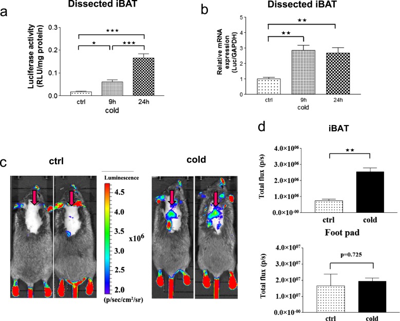
Figure 4.
Cold-induced TH action in the iBAT of THAI #4 mice. (a) luciferaseiferase activity is significantly elevated in dissected iBAT samples of the THAI #4 line after 9 hours at 4°C and further increased after 24 hours of cold stress. *P < 0.05; ***P < 0.001 by one-way ANOVA, followed by Newman-Keuls post hoc test. Means ± SEM (n = 5). (b) Luciferase mRNA increased after 9 hours in dissected iBAT samples of the THAI #4 line; **P < 0.01 by one-way ANOVA, followed by Newman-Keuls post hoc test. Means ± SEM (n = 5). (c) In vivo bioluminescence imaging on iBAT of THAI #4 subjected to cold stress for 24 hours at 4°C, followed by IP luciferin administration. Representative images of two control and two cold-exposed mice. Arrows indicate iBAT. (d) Light intensity diagram of luciferase activity in iBAT and foot pads of control and cold-induced THAI #4 mice. Luciferase activity of skin above the non–BAT-containing region was deducted from iBAT reporter values. Means of photon/s ± SEM (n = 4). **P < 0.001 by Student t test.

