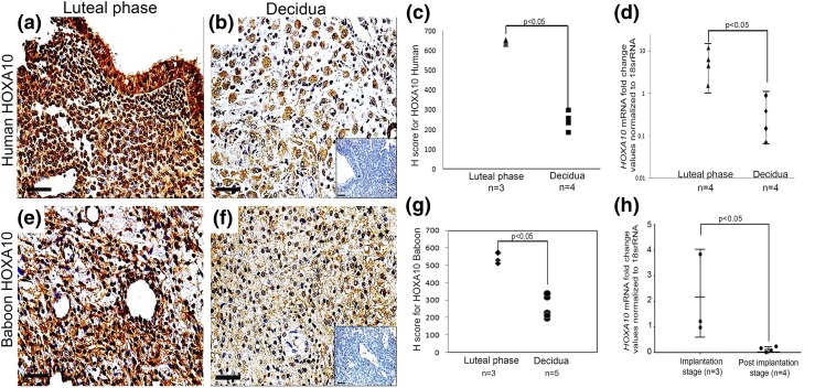
Figure 1.
Expression of HOXA10 in the human and baboon luteal-phase endometrium and in the decidua. Immunohistochemistry for HOXA10 was done on (a) luteal-phase human endometrium, (b) first-trimester human decidua, (e) day 18 baboon endometrium, and (f) day 40 baboon decidua. The respective negative controls without the primary antibody are shown in insets in (b) and (f). The sections are counterstained with hematoxylin. Scale bar, 50 µm. HOXA10 immunostaining was evaluated semiquantitatively (Supplemental Table 2), and the H scores for the biological replicates (n) are represented in (c) and (g) for human and baboon, respectively, where each dot represents one sample. Levels of HOXA10 mRNA were measured by real-time reverse transcription PCR in the (d) luteal-phase human endometrium, first-trimester decidua (10 to 12 weeks of pregnancy), and (h) implantation-stage baboon endometrium (days 20 to 25) and decidua (days 40 to 60). Values on the y-axis are fold change in expression of HOXA10. The number of samples (n) analyzed in each group are given in each graph along with the respective P values.

