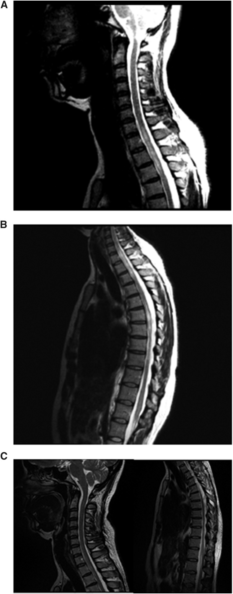
Figure 1.
Imaging of the spinal cord. (A) Magnetic resonance imaging T2 sequences showing hypersignal in the cervical spinal cord C2, C6-C7, and C7-T1. (B) The same widespread change is observed in the thoracic spinal cord, C7-T1, and T9-T10, determining expansion at some levels, suggestive of an inflammatory process. (C) After treatment with methylprednisolone for two cycles, important reduction of lesions in cervical and dorsal spinal cord was noted.

