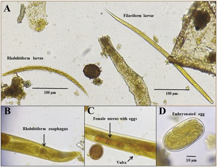
Figure 2.
Photomicrographies of Strongyloides stercoralis stages in fecal smear stained with iodine showing rhabditiform and filariform larva (A), free-living female rhabditiform esophagus (B) and uterus (C), and an embrionated egg (D). This figure appears in color at www.ajtmh.org.

