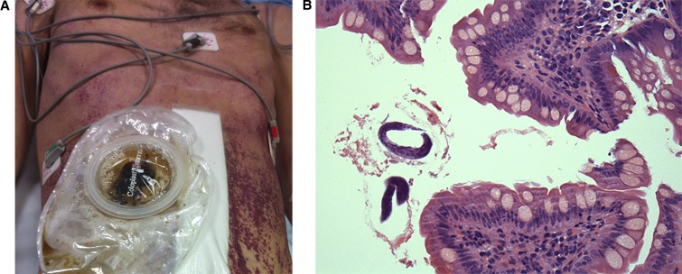
Figure 1.
(A) Patient with ileostomy and purpuric macular abdominal rash characteristic of disseminated strongyloidiasis. (B) Ileal biopsy, magnification ×400, haematoxylin and eosin stain (H&E) stain demonstrating rhabtidiform Strongyloides larvae. This figure appears in color at www.ajtmh.org.

