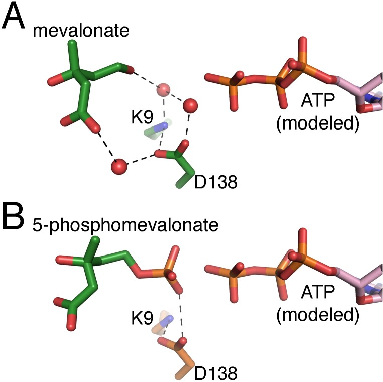
Fig 6. The MmMK active site.
(A) The mevalonate-bound MmMK active site, with mevalonate and MmMK residues in green, and (B) the 5-phosphomevalonate-bound MmMK active site, with 5-phosphomevalonate in green and MmMK residues in orange. ATP modeled from an alignment with the ATP-bound RnMK structure (PDB entry 1KVK) is shown in pink.

