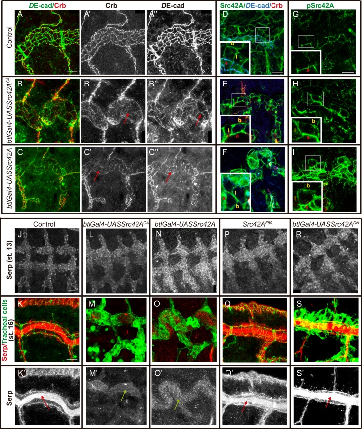
Fig 3. Src42A overactivation affects the accumulation of proteins that undergo endocytic trafficking in the tracheal system.
Lateral views showing a region of the DT of embryos of indicated genotypes stained for the indicated markers. (A-C) Note that Crb accumulation is decreased in the DT (red arrows in B' and C') and DE-cad staining is fragmented and decreased in AJs (red arrows in B'' and C'') in embryos expressing constitutively active Src42A (B) or in Src42A overexpression conditions (C). Scale bar 7,5 μm (D-F) Src42A protein accumulates to cell membranes in control (D) and Src42A overactivation (E) or overexpression conditions (F). Insets (corresponding to close-ups of the regions marked by rectangles) show the accumulation in the apical (a), lateral (l) and basal (b) domains of the membrane. Images show single confocal sections. Scale bar 10 μm (G-I) In contrast to Src42A protein, activated pSrc42A protein accumulates exclusively to the apical region (a) of the membrane in the control (G), but expands along the lateral (l) and basal (b) membrane in Src42A overactivation (H) or overexpression conditions (I). Insets (corresponding to close-ups of the regions marked by rectangles) show the accumulation in the membrane domains. Images show single confocal sections. Scale bar 7,5 μm (J-S) Images show accumulation of Serp (red or white) in tracheal cells (marked in green with DE-cad, K,Q, or GFP in embryos carrying btlGal4-UAS-Src-GFP, M,O,S). Serp is accumulated normally in tracheal cells at early stages in all conditions analysed (J,L,N,P,R). At st 16 Serp is normally accumulated in the luminal compartment associated with the chitin filament (red arrows) in control (K,K') and Src42A loss of function conditions (Q,Q',S,S'). This accumulation is lost (green arrows) in Src42A overactivation (M,M') or overexpression conditions (O,O'). Scale bar J 10 μm, K 7,5 μm.

