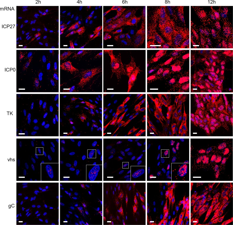
Fig 6. HSV1 transcripts exhibit differential subcellular localisation.
HFFF cells grown in slide chambers were infected with Wt virus at a multiplicity of 2, fixed at 2, 4, 6, 8 or 12 hours after infection, and processed for mRNA FISH with probes to the IE transcripts, ICP27 and ICP0, the E transcript TK and the late transcripts vhs and gC (all in red). Nuclei were counterstained with DAPI (blue). Scale bar = 20 μm.

