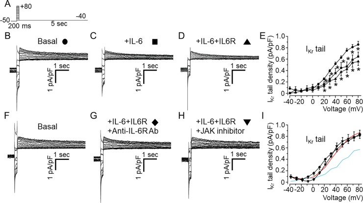
Fig 5. Effects of IL-6 and IL-6+IL-6R on IKr in adult guinea-pig ventricular myocytes.
A, Voltage protocol used for evoking IKr in freshly isolated ventricular myocytes from adult guinea-pig heart. Tail current traces measured in the presence of 100 μM chromanol 293B and 5 μM nifedipine in control untreated cardiomyocyte (B, circle, n = 15), and myocytes pre-treated with IL-6 alone (C, square, n = 8) or IL-6 +IL-6R (D, upward triangle, n = 5). E, Population IKr tail density-voltage curves in basal, IL-6- and IL-6R-treated adult guinea-pig ventricular cardiomyocytes. F, Representative IKr traces in basal condition, (G) pre-treatment with IL-6+IL-6R in the presence of anti-IL-6R antibody (Ab) at 100 μg/ml or, H) a JAK inhibitor-I (5 μM). I, the mean IKr tail density-voltage curves show that the inhibitory effect of IL-6+IL-6R on IKr is reversed in the presence of anti-IL-6R antibody (diamond, n = 12) or JAK inhibitor I (downward triangle, n = 5). For visual comparison, data for control IKr (Red trace) and in the presence of IL-6+IL-6R (Cyan trace) in I.

