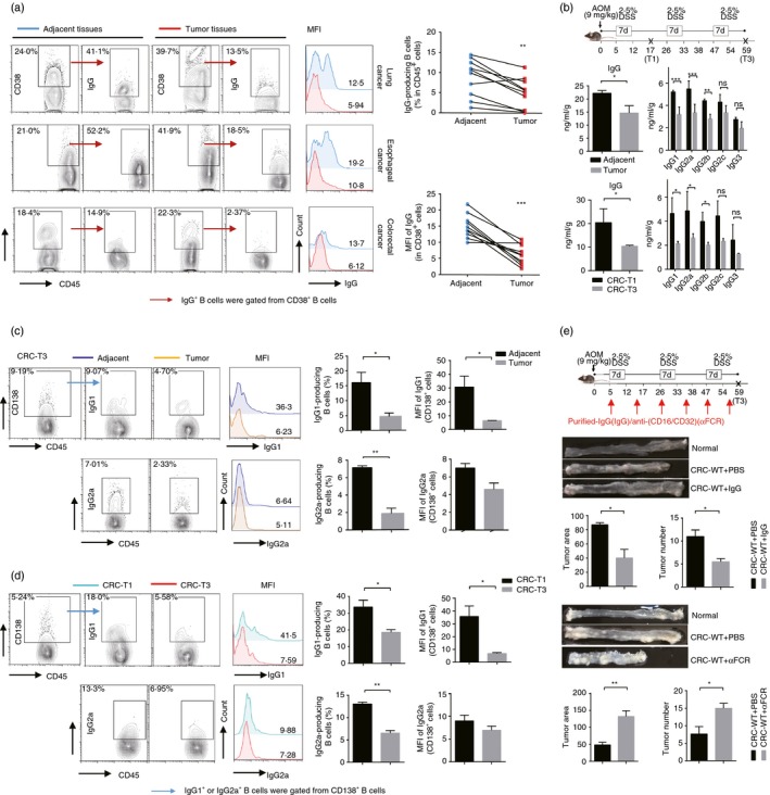
Figure 1.

IgG‐producing B cells mediate anti‐tumor immune response, but decrease in neoplastic tissues during tumor progress. (a) The expression of total IgG including the surface and the intracellular IgG in CD38+ B cells in neoplastic and adjacent normal tissues of human lung, esophageal and colorectal cancers. (b) The concentration of IgG and its subclasses in the adjacent normal tissues and tumor tissues of colorectal cancer (CRC) mice at stage T3 (top), and in the colon tissues from CRC mice at stage T1 and T3 (bottom). (c) Fluorescence‐activated cell sorting (FACS) analysis of IgG1‐producing and IgG2a‐producing B cells, which all gated from CD138+ B cells in adjacent normal sites and tumor sites in the colon tissues from CRC mice at stage T3. (d) FACS analysis of IgG1‐producing and IgG2a‐producing B cells in colon tissues from CRC mice at stage T1 or stage T3. (e) Purified IgG or anti‐CD16/CD32 (α FCR) antibodies were intravenously injected into the C57BL/6 mice at the indicated time‐point, and the CRC model was constructed. Tumor numbers and volumes were detected at stage T3. Mice treated with phosphate‐buffered saline were served as the negative controls. Data shown in (b) to (e) are pooled from three independent experiments each with three or four mice and expressed as mean ± SEM. *P < 0·05, **P < 0·01, ***P < 0·001.
