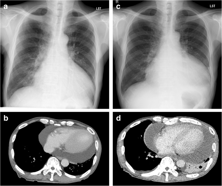
Fig. 1.
Severe cardiomegaly revealed on chest radiography at the initial outpatient appointment (a). Chest computed tomography showed a large pericardial effusion with an estimated volume of 700 mL (b). Cardiomegaly worsened 4 weeks after the initial appointment (c). Chest computed tomography showed a huge pericardial effusion with an estimated volume of 1300 mL (d)

