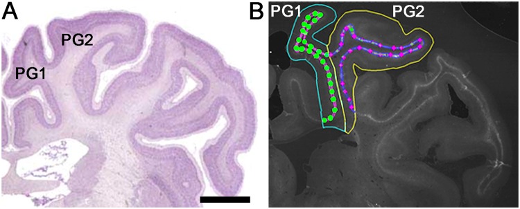
Figure 1.
Sampling regions in the parasagittal cortex of the near-term fetal sheep brain. (A) Sheep brain atlas showing the first (PG1) and second (PG2) parasagittal gyri at the mid-striatal level. (B) Representative tracing and sampling sites of the Wisteria floribunda agglutinin+ (WFA+) layer for PG1 and PG2. The WFA+ layer is traced in pink (PG1) or blue (PG2), while the sampling sites are marked with circles (PG1) or diamonds (PG2). (A) was adapted with permission from http://www.brains.rad.msu.edu, supported by the US National Science Foundation and the National Institutes of Health. Scale bar: 5 mm.

