Figure 3.
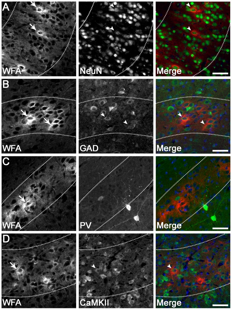
PNN expression on various neuronal subtypes in the parasagittal cortex. Dense pericellular WFA reactivity (arrows) was observed around subsets of NeuN+ neurons (A; arrowheads), GAD+ interneurons (B; arrowheads), and CaMKIIα+ excitatory neurons (D; arrowheads), in a pattern resembling immature PNNs. By contrast, very few PNNs (arrows) were localised to parvalbumin (PV)+ interneurons (C). Note that the diffuse extracellular component of WFA staining is observed within layer 6 (dotted lines). Scale bar: 50 µm.

